Hernia of the cervical spine is an occupational disease of athletes and people whose work activity is associated with weight lifting and vibration. With this pathology, there is a rupture of the fibrous ring of the intervertebral disc located in the cervical spine, as a result of which it loses the possibility of amortization.
Features:
The neck is the upper part of the spinal column, which is characterized by high mobility, allowing free and varied head movements. It consists of 7 vertebrae with transverse processes, on either side of which are blood vessels and spinal nerves. The two upper vertebrae of the neck differ from others in anatomical structure. They connect the spine to the skull. Between the paired adjacent vertebrae, there are intervertebral discs, consisting of an annulus fibrosus and a nucleus pulposus pulposus.
A hernia is formed mainly between 5 and 6 discs, as well as 6 and 7 cervical vertebrae. Much less often, the disease affects the space between the 4th and 5th vertebrae of the neck. Almost never, pathology occurs between the 7 cervical and 1 thoracic vertebrae.
The occurrence of prolapse provokes ring rupture and disc protrusion. The compression of the spinal roots is manifested by a sharp pain syndrome. Due to the close location of the arteries of the spinal section, a hernia can cause neurological disorders and vascular pathologies.
The size of the neck vertebrae is much smaller than that of the thoracic and dorsal. However, the anatomical features of this area are such that even the slightest protrusion can provoke the appearance of a hernia.
Types and stages
The discs may be in a state of pre-hernia or true prolapse. There are several stages of the disease, each of which has characteristic features:
- the first - the intervertebral disc is intact, the size of the protrusion does not exceed 0.2 cm;
- the second - there is damage to the annulus fibrosus, the degree of protrusion exceeds 0.2 cm and can reach 0.4 cm;
- the third - there is a rupture of the ring and a strong displacement of the disk up to 0.6 cm;
- the fourth is a critical degree of damage that threatens the development of sequestration. The dimensions of the prolapse at this stage reach 0.8 cm.
Sequestration is a complicated form of hernia, which consists in the final detachment of a deformed fragment of cartilage from the disc and getting it into the space of the spine.
The danger of this condition lies in the possibility of the rapid development of serious damage to the nerve endings of an irreversible nature and their death. There is a high risk of paralysis of the trunk below the affected area, partial or complete paresis of the hands, dysfunction of the reproductive system and urogenital organs.
The reasons
A healthy person does not experience discomfort and pain when bending and turning the neck. Degenerative processes reduce nutritional levels and disc amortization.
The reasons for the development of this pathology are:
- spinal injury;
- hypodynamia;
- improper posture;
- osteochondrosis.
People with a genetic predisposition to hernia are subject to the accelerated development of pathological changes. In addition, the increase in the rate of degeneration processes is influenced by age-related changes, the presence of other congenital defects and unfavorable working conditions.
Symptoms
Acute pain syndrome in the shoulder joints, radiating to the head and neck, a state of numbness and limited mobility of the limbs are the main signs that allow diagnosing a hernia of the cervical spine. Tilting the neck increases pain. The presence of this pathology can provoke brain hypoxia.
For a hernia, the following symptoms are characteristic:
- the occurrence of dizziness;
- violations of gait and coordination of movements;
- drops in blood pressure;
- short-term fainting;
- sudden darkening in the eyes.
Pathology has a variable clinical picture, depending on the area of the lesion.
| Location | Signs |
| C2-C3 | Migraine, loss of sensitivity of the tongue, sore throat, difficulty turning the head, decreased vision. |
| C3-C4 | Soreness in the clavicle, discomfort when lifting shoulders and head movements, migraines. |
| C4-C5 | Localization of pain in the area of the forearm muscles. Raising your arms above your head increases the discomfort. |
| C6-C7 | Decreased muscle tone in the triceps, thumb and forearm. Tingling sensation on the skin. |
| C7 and 1 thoracic region | Weakness and limited movement of the hand, the possibility of pain spreading throughout the hand. |
Diagnostics
The presence of the above symptoms is a reason for visiting a neurologist. The specialist will conduct a study of reflexes and sensitivity in the upper limbs and shoulders, find out the localization of the pathology and prescribe a thorough diagnosis.
There are several methods for detecting the presence of a hernia:
- radiography;
- CT;
- MRI;
- myelogram.

MRI scan of the cervical spine. © Maxim Pavlov - stock.adobe.com
Treatment
After a thorough examination of the patient, the neuropathologist chooses the appropriate treatment regimen for him. He must determine whether it is possible to use non-surgical methods of treating a herniated cervical disc or whether a neurosurgeon's examination is necessary.
In the absence of an obvious violation of cerebral circulation, there is no need for surgical intervention.
If drug treatment does not give an effect within six months or the patient's condition worsens, the council of neurosurgeons decides on the operation.
Conservative therapy is based on the principles:
- improving the nutrition of the annulus fibrosus of the damaged disc;
- relaxation of the neck muscles;
- strengthening the volume of the cervical muscles to fix the neck;
- getting rid of pain that prevents the vertebrae from being in a normal position.
The current types of treatment for this pathology will be discussed below.
Mode
During the first week, the patient should use a Shants collar or other fixation orthoses, or stay in bed. This allows the diseased disc to recover and take in the nucleus pulposus.

Shants collar. © mulderphoto - stock.adobe.com
Removal of the device is allowed after the pain in the arms and shoulders has faded. Initially, the retainer is removed during sleep, then - for taking hygiene procedures. When the patient's condition improves and there is no pain, the collar is removed for the whole day. You can't twist your head or stretch your neck.
It is recommended to take a shower for the entire period of treatment, since in the bathroom a person is in a position that is not physiological for the neck.
Drug treatment
Neck hernia therapy involves the use of such drugs:
- Anti-inflammatory. Designed to eliminate painful sensations. First, they are prescribed in the form of injections, at the second stage of treatment, they can be taken in tablet form.
- Muscle relaxants. They are used to relieve spasm and relax skeletal muscles. Initially, intramuscular injections are prescribed, and then tablets.
- Chondroprotectors. The regeneration of the annulus fibrosus is started. Applied for at least 6 months. In the presence of severe weakness, a burning sensation or numbness in the hand, it is possible to block the affected segment of the vertebral region by using a combination of novocaine and glucocorticoids. The frequency of use of these drugs should not exceed 4 times within two months.
Physiotherapy methods
Physiotherapy is used after the acute phase of the disease has been removed and in the elimination of pain. The following methods are used:
- diadynamic therapy;
- paraffin applications;
- electrophoresis with novocaine;
- magnetotherapy;
- ozokerite applications on a sore spot.
Massage
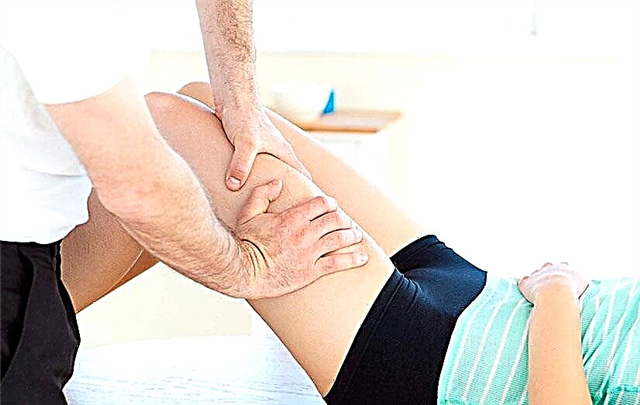
The procedure must be performed with the utmost care by a suitably qualified person. The task of the masseur is to relieve spasm and normalize muscle tone. The main thing is not to provoke pinching of the vertebral arteries or spinal cord.

© WavebreakmediaMicro - stock.adobe.com
Manual therapy
Before proceeding with the procedure, the chiropractor should become familiar with the patient's MRI or CT scans. The provided research results allow the specialist to navigate where his efforts should be directed to restore the spine.
Physiotherapy
The type of exercise therapy for neck prolapse is selected depending on the period of the disease. Effective gymnastics techniques were developed by doctors Bubnovsky and Dikul. During the acute phase, only diaphragmatic breathing exercises are allowed in the supine position.
At the end of the first week, emphasis should be placed on strengthening the muscles of the upper limbs:
- circular rotation with brushes;

- circular rotation in the elbow joints, their flexion and extension.


- clenching and unclenching of fists.

After another two weeks, it is recommended to use neck exercises that help strengthen the muscular corset:
- Lying on your back, apply pressure alternately with the back of your head on the couch and your forehead on the assistant's palm.
- Lying on your stomach, pressure first with your forehead on the couch, and then with the back of your head on the doctor's palm.
- From a sitting position, alternate pressure on the arm with the forehead and the back of the head. The same can be done from a standing position.


- While standing, the shoulders are raised and lowered. The same can be done while sitting on a chair with your palms on the table.

- Starting position is sitting on a chair, hands on knees. Gentle turns of the head left and right with a delay of 5 seconds. (10 times each side).

A set of four exercises:
- Standing, back straight, arms along the body. Gently tilting the head back with a deep breath and tilting the head down with the chin towards the chest with exhalation (10 times).
- The same starting position. Circular head movements in both directions (10 times).
- Head tilts to the left (10 times).
- The same movement to the right (10 times).

Other exercises:
- Regular pull-ups on the horizontal bar. You should start at 5 times a minute, gradually increasing the amount to 10.

- Push-ups from the floor (6 times).

Exercises for a herniated disc should be performed in the morning.
After gymnastics, it is better not to go outside. This will avoid hypothermia, which is harmful to the spine. The duration of rehabilitation is determined by the doctor and depends on the effectiveness of the treatment. If you experience discomfort and pain, you should stop exercising.
Hirudotherapy
A treatment method based on the healing properties of medicinal leeches. Their saliva has a high content of hirudin. It improves blood circulation in the area of the damaged cervical vertebra and prevents blood clots. During the bite, leeches suck out up to 15 ml of blood. In this case, peptidase, hirustazine and collagenase enter the human body.

© 2707195204 - stock.adobe.com
Vacuum therapy
This technique is familiar to many under the name of cupping massage. It is of two types:
- Static. Banks are placed along the spine for 15-20 minutes.
- Dynamic. The doctor moves the containers along the patient's back, which has been previously lubricated with cream or oil.
The procedure activates metabolic processes, improves blood circulation and eliminates congestion.
Plasma therapy
Regenerative medicine technique based on the patient's blood plasma. During the preparation process, hormone-like polypeptides are released from platelets, which can accelerate the process of tissue repair.
Blood is drawn initially. The test tube with the obtained biological fluid is placed in a centrifuge to produce plasma. The finished product is injected into the affected segment of the spine by injection.
Additional therapies
In addition to the listed methods of treatment, acupuncture and the method of post-isometric relaxation are also used - these are special exercises that are performed in conjunction with an exercise therapy specialist.
Operative treatment
Surgical intervention is planned for:
- the presence of signs of impaired cerebral circulation: dizziness, headache, decreased sense of smell, hearing and vision;
- lack of effect from conservative methods of treatment;
- identification of large sequesters in the spinal canal.
There are three ways to remove a hernia:
- Anterior discectomy and osteosynthesis. The surgeon makes an incision in the front of the neck, about 3 cm long. After removing the damaged part of the disc, the vertebrae are fused together with or without a bone graft.
- Posterior discectomy. This involves making an incision in the back of the neck. With the help of a gauze tampon clamped in tweezers, the doctor pushes the muscles aside and exposes the bone tissue of the vertebral process. A portion of the bone is removed to allow access to the disc and retrieval of the hernia. At the end of the surgery, the nerve roots are no longer clamped.
- Microendoscopic discectomy. This is a minimally invasive operation. Access to the damaged area of the spine is performed from the back of the neck. All medical manipulations are carried out with small instruments. The operation is performed under endoscopic control.
Complications
Late diagnosis of the disease can adversely affect health and provoke the following consequences:
- scoliosis;
- violation or cessation of breathing due to damage to the spinal cord;
- muscle weakness in the arms, including complete or partial paralysis;
- decreased hearing and vision;
- neurotic disorders;
- disruption of the digestive tract;
- frequent fainting;
- low circulation of blood flow in the brain and spinal regions.
The listed complications are extremely dangerous. Some of them require urgent medical attention. They can significantly reduce the quality of human life and cause death. It is very important to diagnose the disease in a timely manner.
In the early stages, a hernia of the cervical spine is effectively amenable to therapeutic correction. As a preventive measure, it is recommended: adherence to the correct diet, visit the pool, play sports, avoid hypothermia and intense physical exertion on the cervical spine.


